2 D Echocardiography with Colour Doppler & Strain Imaging
What is 2D Echocardiography with Colour Doppler & Strain Imaging?
It is an advanced, non-invasive heart imaging test that combines standard 2D echocardiography with colour Doppler flow analysis and strain imaging. This test allows doctors to visualize the heart’s chambers, valves, and blood flow in real-time while assessing the function and subtle motion of the heart muscles.
Why is This Test Important?
2D Echocardiography with Colour Doppler & Strain Imaging is a powerful combination that provides a detailed, three-dimensional perspective of heart health. Traditional 2D echo allows cardiologists to assess the size, shape, and motion of the heart’s chambers and valves. It helps in identifying issues like enlarged chambers, thickened heart walls, or valve dysfunction.
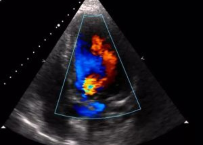
The Colour Doppler component adds a visual layer showing blood flow in real-time. It displays the speed and direction of blood movement using color-coded images, which is critical in detecting turbulent or reverse blood flow, indicating problems such as valve leakage (regurgitation), narrowing (stenosis), or congenital defects like septal holes.
What makes this test even more valuable is Strain Imaging—an advanced method to detect subtle abnormalities in how the heart muscle contracts and relaxes. It quantifies the deformation (or “strain”) of the heart walls with precision. This is particularly useful in identifying early myocardial damage in patients who may not show any symptoms yet—such as those undergoing chemotherapy, with early heart failure, or with hypertension and diabetes.
Together, these techniques allow for earlier detection, better risk stratification, and more accurate monitoring of heart disease. The test guides treatment plans, tracks disease progression, and evaluates the effectiveness of therapies—making it an essential tool in modern cardiology.
- Detects early heart muscle damage.
- Tracks heart changes in high-risk patients.
- Assesses valve function and blood flow.
- Helps monitor heart during chemotherapy.
- Provides detailed insight into heart motion.
- Safe, non-invasive, and highly accurate.
Head of Department, Cardiology Services, Suburban Diagnostics India Private Limited.
Make Appointment
- 3 PM - 5 PM , Monday - Friday
Copyright @2025. All Right Reserved | Designed By Rebecca Digital